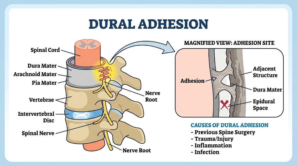
Dural adhesion refers to the abnormal binding of the dura mater—the outermost protective membrane of the spinal cord—to surrounding structures due to scar tissue formation. Typically arising from spinal surgery, trauma, or inflammation, these fibrous bands can obliterate the epidural space and tether nerve roots. This restriction of normal nerve mobility often leads to chronic pain, neurological deficits, and complications during subsequent spinal procedures, making targeted management and prevention critical.
1. Introduction
Dural adhesion (DA) refers to abnormal fibrotic sticking between the dura mater and surrounding tissues, such as the ligamentum flavum, arachnoid, or postoperative scar tissue. This adhesion may restrict neural mobility, distort dural tension, and produce neurological symptoms. DA is increasingly recognized following trauma, inflammation, surgery, or structural abnormalities. Recent biomaterial and regenerative research highlights the complexity of preventing adhesions and restoring healthy dural mechanics.1,2,3
2. Etiology
Dural adhesion arises from multiple biological and mechanical processes. Inflammatory conditions such as arachnoiditis cause leptomeningeal thickening, fibrotic tissue formation, and nerve root clumping, which lead directly to dural adhesions.4 Postsurgical scarring is another major factor: following spinal surgery, fibroblasts proliferate between the dura and surrounding tissues, forming dense adhesions that impair mobility and increase the risk of reoperation.
Biomechanical factors also contribute. Research on patients with ossification of the ligamentum flavum (OLF) shows that dural ossification and adhesion commonly coexist and increase the likelihood of intraoperative dural tears during decompression procedures.6 Additionally, structural abnormalities such as dural ectasia produce weakened or enlarged dura, predisposing the tissue to abnormal adherence.5
3. Symptoms
Dural adhesion can manifest with a wide spectrum of symptoms depending on location and severity:
- Deep axial spine pain or stiffness
- Neuropathic symptoms such as numbness, tingling, or burning
- Leg or arm heaviness or fatigue during motion
- Pain aggravated by spinal flexion, extension, or coughing
- Restriction of dural mobility leading to tension-induced pain
- In severe cases, gait disturbance or functional impairment
Patients with adhesive arachnoiditis often experience progressive neurological decline due to compressive fibrosis and nerve root tethering.4
4. Diagnosis
Diagnosis involves a combination of history, examination, and imaging:
- MRI is the primary imaging tool to detect nerve root clumping, scarring, and reduced cerebrospinal fluid spaces.
- CT is useful in identifying ossified structures associated with adhesion, such as in OLF cases.6
- Neurological tests assess dermatomal sensory changes, motor deficits, and dural tension signs.
- Osteopathic evaluation identifies motion restriction consistent with tethered dura, including dural tension during cervical or lumbar flexion.
5. Treatment
5.1 Conventional Medical and Surgical Approaches
Research continues to refine biomaterial-based strategies for dural repair. Janus SIS membranes, hyaluronic acid–CMC hydrogels, and wet-adhesive bilayer patches demonstrate potential to reduce postsurgical adhesion formation and promote healing following dural injury.1,2,3 These technologies aim to prevent the cycle of fibrosis, inflammation, and mechanical restriction.
Surgery may be indicated for severe cases involving:
- Dural tears with cerebrospinal fluid leakage
- Severe arachnoiditis with neurological compromise
- Adhesive fibrosis causing nerve root compression
5.2 Osteopathic Manual Treatment (OMT)
OMT is frequently used to improve dural mobility and reduce tension. Key principles include restoring balance to the cranium, spine, sacrum, and dural tube through gentle, indirect techniques.
Common osteopathic techniques:
- Craniosacral therapy: Balances dural tension and enhances cerebrospinal fluid dynamics.
- Balanced ligamentous tension (BLT): Applied around the lumbosacral junction to reduce strain on dural attachments.
- Indirect sacral technique: Releases tension near the filum terminale.
- Cervical dural release: Reduces neural tension during flexion and rotation.
- Thoracic inlet release: Improves venous and lymphatic drainage to reduce inflammatory congestion.
By reducing abnormal strain and improving dural gliding, OMT may lessen symptoms related to mechanical tethering.
5.3 Home Care and Self-Management
1. Gentle Neural Mobilization
Slow, controlled nerve glide exercises reduce mechanical tension without provoking inflammation.
2. Posture Optimization
Neutral spine positioning decreases dural stretch and irritation, especially during prolonged sitting or bending.
3. Heat Therapy
Warm compresses relieve muscle tension surrounding dural attachments.
4. Movement Hygiene
Patients should avoid repetitive spinal flexion or heavy lifting during flare-ups.
Consistent home management can complement clinical interventions and improve long-term outcomes.