Superior gluteal nerve entrapment syndrome (SGNES) is a rare but clinically significant cause of buttock, hip, and lower-limb dysfunction. Although less commonly recognized than sciatic or cluneal nerve entrapment, its impact on gait, lumbopelvic stability, and chronic gluteal pain is substantial. Early literature first described SGNES as a neuropathy caused by muscular or tendinous compression—most notably involving the piriformis and surrounding structures1. Subsequent anatomical and clinical studies have confirmed that the superior gluteal nerve (SGN) can be compressed within the suprapiriformis foramen, producing characteristic patterns of pain, weakness, and functional loss2.
Etiology
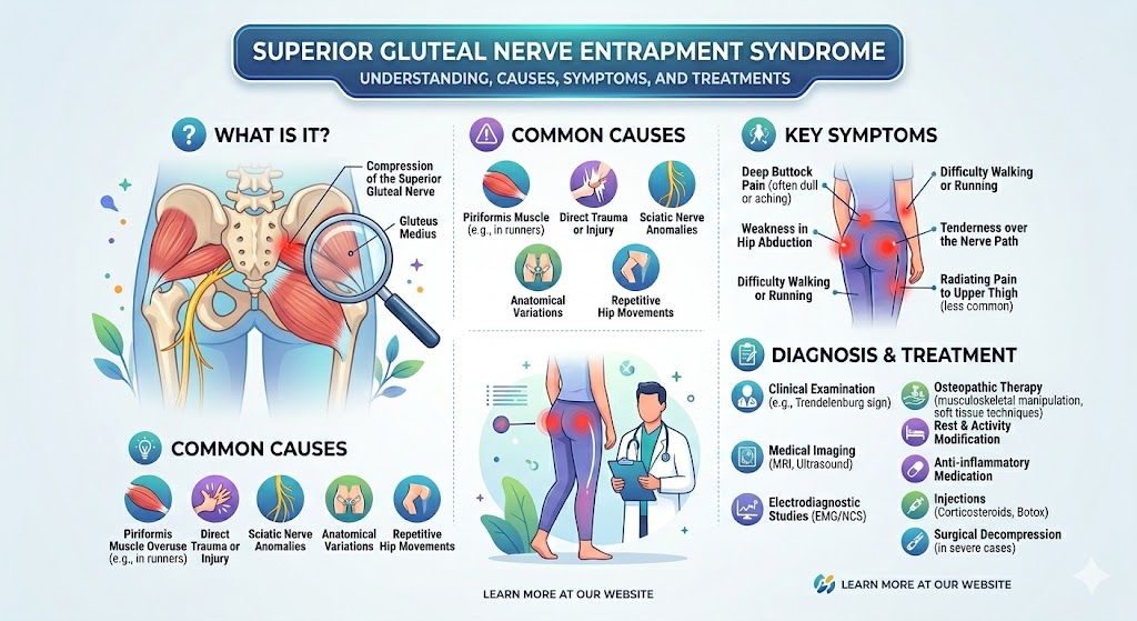
The SGN arises from the L4–S1 roots and exits the pelvis through the suprapiriformis foramen. It innervates the gluteus medius, gluteus minimus, and tensor fasciae latae—key stabilizers of pelvic alignment and gait. Entrapment may occur anywhere along this pathway. Early descriptions identified compression by the anterior-superior tendinous fibers of the piriformis muscle as a common cause1. A large anatomical dissection study later demonstrated that variations in the suprapiriformis foramen shape, fibrous bands, or aberrant muscle slips can significantly narrow the nerve’s passageway, predisposing it to irritation or chronic compression2.
Additionally, deep gluteal syndrome (DGS)—a broader category of nerve compression disorders within the deep gluteal space—can involve the SGN, though it more commonly affects the sciatic nerve. DGS manifests as posterior hip pain due to compression of peripheral nerves by muscles, fibrous bands, or vascular structures4. SGNES represents a more specific subset focused on the SGN but may coexist with other neuropathies.
Biomechanical dysfunction and repetitive stress can also contribute. Chronic hip abductor overuse, gluteal tendinopathy, lumbopelvic instability, or compensatory gait patterns place tension along the SGN pathway. Rarely, traumatic events, previous hip injections, or local scarring may create fibrotic entrapment. Electrophysiological case reports further confirm neuropathy patterns associated with chronic nerve compression in the gluteal region9.
Symptoms
SGNES produces a unique constellation of symptoms due to the SGN’s pure motor function and its critical role in hip abduction and pelvic stabilization. Early descriptions emphasized aching, claudication-type buttock pain, often worsened by walking, prolonged standing, or single-leg stance7. Patients frequently report weakness when lifting the leg sideways, a sense of pelvic dropping, or instability during gait.
Typical symptoms include:
– Deep, aching lateral or posterolateral buttock pain
– Pain radiating toward the lateral hip or sacroiliac region
– Fatigue or heaviness in the gluteal muscles after walking
– Trendelenburg gait or difficulty maintaining level pelvic alignment
– Difficulty standing on one leg
– Worsening pain with hip abduction or internal rotation
– Relief with rest or unloading the affected hip
Unlike sciatic neuropathy, SGNES usually produces no distal paresthesia. However, chronic compensatory movement patterns may lead to secondary issues, including low-back strain, contralateral hip overuse, or development of cluneal nerve irritation. Related literature on superior cluneal nerve entrapment underscores the frequency of misdiagnosis in gluteal and lumbar pain syndromes, emphasizing the need for careful differentiation5.
Diagnosis
Diagnosis relies on clinical assessment, supported by imaging and electrodiagnostic testing when necessary. Provocative maneuvers that stretch or contract the gluteus medius and minimus—such as resisted abduction testing—typically reproduce pain. Significant weakness in hip abduction or a positive Trendelenburg sign strongly suggest SGN involvement.
Electromyography can detect denervation or neuropathic changes in SGN-innervated muscles, helping differentiate SGNES from lumbar radiculopathy or superior cluneal nerve entrapment9. Ultrasound and MRI may identify muscle atrophy, tendon abnormalities, or impingement caused by anomalous structures. Anatomical variation studies indicate that the SGN may innervate the piriformis muscle in up to 70% of individuals, complicating interpretation of symptoms and reinforcing the importance of anatomical awareness10.
Treatment Approaches
Conservative Management
Conservative treatment is the first-line approach. A systematic review found both conservative and surgical methods effective for deep gluteal syndrome, though high-quality comparative evidence remains limited6. Conservative care generally includes:
– Activity modification
– Targeted physiotherapy
– Manual therapy or osteopathic interventions
– Anti-inflammatory modalities
– Image-guided injections (when indicated)
Traditional Chinese medicine approaches, such as needle-in-row therapy combined with herb-partition moxibustion, have demonstrated symptom reduction and functional improvement in clinical observation studies3.
Osteopathic Treatment
Osteopathic manipulative treatment (OMT) focuses on restoring mobility, reducing muscular imbalance, and decreasing compressive load on the SGN. Techniques may include:
– Myofascial release to piriformis, gluteus medius/minimus, and TFL
– Muscle energy techniques to correct pelvic rotations or sacroiliac dysfunction
– Counterstrain for gluteal trigger points
– Balanced ligamentous tension for sacroiliac and lumbopelvic mechanics
– Articulatory techniques to improve hip internal rotation and abduction
OMT aims to open the suprapiriformis space, reduce tension on the nerve pathway, and restore balanced gluteal activation. Addressing thoracolumbar junction mobility is often essential, as compensation patterns in this region can contribute to lumbopelvic misalignment.
Surgical Treatment
Surgical decompression is rare but may be necessary when conservative care fails. Procedures focus on releasing fibrous bands or abnormal structures causing compression. Outcomes are generally favorable when careful diagnostic criteria confirm true SGNES.
Home Care and Self-Management
Home care supports long-term recovery and complements manual therapy. Key components include:
1. Gluteal Strengthening
Targeted strengthening of the gluteus medius and minimus improves pelvic control and reduces nerve tension. Exercises include side-lying abduction, standing hip abduction with resistance, and controlled single-leg stance practice.
2. Flexibility and Release Work
Gentle stretching of the piriformis, TFL, and iliotibial band reduces compressive forces around the suprapiriformis foramen.
3. Posture and Ergonomics
Neutral pelvic alignment during walking and standing is critical. Patients should avoid prolonged sitting on firm surfaces and reduce repetitive hip adduction activities that irritate the nerve.
4. Heat and Self-Massage
Heat therapy and soft tissue self-massage using a foam roller can alleviate muscle tension and improve circulation.
With consistent home care and appropriate clinical intervention, most individuals experience substantial improvement in symptoms, gait, and functional stability.