1. Introduction: A Living Tissue Without Blood Vessels
Articular cartilage is the smooth, resilient tissue covering the ends of bones in synovial joints, including the knees, hips, shoulders and ankles. It helps distribute mechanical loads and allows opposing joint surfaces to move with minimal friction. Although cartilage appears relatively simple, it is a highly organized biological tissue whose structure is essential to joint function.
A distinctive feature of mature articular cartilage is that it is avascular: it contains no conventional network of blood vessels. It also lacks lymphatic vessels and has no direct nerve supply within the cartilage itself. Consequently, its resident cells, called chondrocytes, cannot obtain oxygen and nutrients through the same direct vascular route used by many other tissues. [3] [4]
Instead, cartilage depends on the movement of molecules through its surrounding environment and dense extracellular matrix. This arrangement allows cartilage to withstand repeated loading, but it also limits its capacity for repair. Understanding how nutrients reach chondrocytes helps explain the relationship between joint movement, cartilage biology and long-term joint health.
2. Synovial Fluid: The Main Nutrient Source
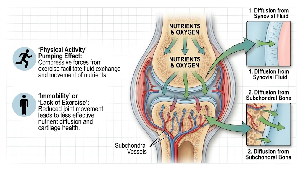
In adult synovial joints, synovial fluid is generally considered the principal source of nutrients for articular cartilage. This fluid is produced and regulated by the synovial membrane and contains water, electrolytes, dissolved gases, glucose, amino acids and other molecules required for normal joint function. It also contains hyaluronan and lubricating molecules that contribute to the fluid's viscoelastic and boundary-lubricating properties. [3] [5]
Synovial fluid does more than lubricate the joint. It provides a medium through which oxygen and small nutrient molecules travel toward cartilage cells. Metabolic by-products, including lactate, can move in the opposite direction toward the joint fluid, where they can be cleared through surrounding tissues and circulation.
The nutrient supply depends on the composition and condition of the synovial fluid, the distance molecules must travel, and the physical and chemical properties of the cartilage matrix. Healthy synovial fluid and an intact cartilage surface therefore form an important part of the joint's nutritional environment.
3. Diffusion: How Nutrients Enter Cartilage
Diffusion is the movement of molecules down a concentration gradient. When oxygen or a nutrient is more concentrated in the synovial fluid than in nearby cartilage, molecules can move across the cartilage surface and through its extracellular matrix. They eventually reach chondrocytes, which use them to maintain cellular metabolism and produce the components of cartilage tissue. [6]
The extracellular matrix is composed mainly of a collagen network, proteoglycans and water. Collagen provides tensile strength, while proteoglycans attract and retain water, helping cartilage resist compression. This matrix is not an empty sponge: its molecular structure, charge and permeability influence which substances can enter, how quickly they travel, and how deeply they penetrate.
Small molecules such as oxygen and glucose generally move more readily than many larger molecules. Molecular size, electrical charge, shape and interactions with the matrix all affect transport. These properties help explain why cartilage nutrition is a regulated physical process rather than the unrestricted flow of nutrients into tissue. [6] [7]
Chondrocytes use the available oxygen and nutrients to maintain and renew the extracellular matrix. Because oxygen availability is relatively limited in cartilage, these cells rely substantially on anaerobic metabolism. Their metabolic activity is also influenced by mechanical loading, age, inflammation and the condition of the surrounding matrix. [3] [4]
4. Why Movement and Loading Matter
Diffusion occurs even when a joint is still. However, normal joint movement changes the mechanical environment and can assist the exchange of fluid and dissolved substances between cartilage and synovial fluid.
During loading, cartilage deforms and its interstitial fluid pressure rises. Some fluid is displaced from the matrix. When the load is reduced, the tissue partially recovers its shape and fluid can move back into it. Repeated loading and unloading therefore contribute to fluid exchange and can enhance the transport of certain solutes. Researchers describe the relevant processes in terms of diffusion, convection and poroelastic behaviour. [6] [7]
This mechanism is sometimes informally described as a nutrient-pumping effect. The phrase can be useful as a simplified illustration, but it should not be interpreted as a literal pump or a guarantee that exercise will deliver a fixed quantity of nutrients to every cartilage cell. Diffusion remains important, and the effects of loading vary with the tissue, the molecule and the loading pattern.
Appropriate movement also supports muscle strength, joint coordination and general mobility. Conversely, prolonged immobilization may alter the mechanical and metabolic environment of cartilage. These findings support regular, graded movement as part of general joint care, provided the activity is appropriate for the individual's condition. They do not mean that painful or heavily loaded joints should be forced through symptoms.
5. Does Nutrient Supply Also Come from Bone?
Articular cartilage lies directly above subchondral bone, which provides structural support and has a rich blood supply. Nutrient exchange between bone and cartilage has been investigated extensively. However, the importance of this route in mature healthy cartilage is more complicated than a simple direct supply from bone.
The calcified cartilage zone and subchondral plate form an interface that restricts exchange. Experimental findings suggest that subchondral pathways may contribute under some circumstances, but the relative importance of bone-derived nutrients varies with age, species, tissue condition and the methods used to study transport. Synovial fluid remains a major source of nutrition for mature articular cartilage. [1] [2]
Changes in the subchondral bone and cartilage can occur together in osteoarthritis. These changes may affect the local transport environment, but osteoarthritis is a multifactorial condition involving cartilage, bone, synovium and other joint structures. It should not be explained solely as a failure of cartilage nutrition.
6. Why Cartilage Nutrition Matters for Joint Health
Chondrocytes maintain cartilage by synthesizing and regulating the extracellular matrix while also responding to mechanical and biochemical signals. If the matrix is damaged, its structure and permeability can change. Inflammatory mediators and enzymes may accelerate matrix breakdown, while the cells' ability to restore the tissue is limited.
Cartilage has a relatively poor intrinsic repair capacity because it lacks blood vessels and has a low density of cells. Severe damage therefore cannot usually be repaired simply by increasing movement or improving nutrient intake. Cartilage health depends on a balance of cellular metabolism, matrix maintenance, mechanical loading and the surrounding joint environment. [4] [8]
For general joint health, practical measures include maintaining regular, tolerable physical activity; progressively strengthening the muscles that support a joint; avoiding sudden increases in training load; and seeking appropriate assessment for persistent swelling, locking, instability or pain. Diet supports overall health, but no particular food or supplement has been established as a reliable way to regenerate lost articular cartilage.
7. Conclusion
Articular cartilage is a specialized living tissue that receives nutrients mainly through diffusion from synovial fluid rather than through its own blood vessels. Movement and alternating loading and unloading can support fluid exchange, while chondrocytes use available nutrients to maintain the cartilage matrix and its mechanical properties.
Understanding this biology highlights the value of appropriate movement and a healthy joint environment. It also clarifies an important limitation: normal movement supports cartilage function, but it does not guarantee prevention or reversal of degeneration. Long-term joint health depends on several interacting biological, mechanical and clinical factors.
References
- Wang, Y., et al. (2013). Nutrition and degeneration of articular cartilage. Knee Surgery, Sports Traumatology, Arthroscopy, 21, 1751–1762. https://doi.org/10.1007/s00167-012-1977-7
- Arkill, K. P., & Winlove, C. P. (2008). Solute transport in the deep and calcified zones of articular cartilage. Osteoarthritis and Cartilage, 16, 708–714. Search bibliographic record
- Fox, A. J. S., Bedi, A., & Rodeo, S. A. (2009). The basic science of articular cartilage: Structure, composition, and function. Sports Health, 1(6), 461–468. Read the published review
- Hunziker, E. B. (1994). Articular cartilage structure and function. Osteoarthritis and Cartilage, 2, 13–25. Search bibliographic record
- Hall, J. E., & Hall, M. E. (2021). Guyton and Hall Textbook of Medical Physiology (14th ed.). Elsevier. Publisher: Elsevier
- DiDomenico, C. D., Lintz, M., & Bonassar, L. J. (2018). Molecular transport in articular cartilage—what have we learned from the past 50 years? Nature Reviews Rheumatology, 14, 393–403. https://doi.org/10.1038/s41584-018-0033-5
- Lai, W. M., Hou, J. S., & Mow, V. C. (1991). A triphasic theory for the swelling and deformation behaviors of articular cartilage. Journal of Biomechanical Engineering, 113(3), 245–258. https://doi.org/10.1115/1.2894880
- Buckwalter, J. A., & Mankin, H. J. (1997). Articular cartilage: Degeneration and osteoarthritis, repair, regeneration, and transplantation. Instructional Course Lectures, 47, 487–504. Search bibliographic record
- Maroudas, A. (1975). Biophysical chemistry of cartilaginous tissues with special reference to solute and fluid transport. In studies of cartilage swelling and compressive stress. Nature, 260, 808–809. Search bibliographic record
- Basser, P. J., Schneiderman, R., Bank, R. A., Wachtel, E., & Maroudas, A. (1998). Mechanical properties of the collagen network in human articular cartilage as measured by osmotic stress technique. Archives of Biochemistry and Biophysics. Search bibliographic record